
Following the review of the initial PIS modules, Gill (with input from Giovanna) compiled a list of potential illustrative images for use in the modules. Some of these will depict equipment used to take samples and conduct the actual imaging (similar in style to the header image of this post). Other images will show study participants and their role in the study. Those illustrations will probably be similar in style to images Gill created to represent ‘Executive Function’, in another project for the CNS (examples can be seen on Gill’s website).
The initial list is as follows, although this will undoubtedly change and develop as the project progresses.
| Type of image | Module |
| Person in an MRI scanner | 2, 4, 5, 10 |
| Person filling in questionnaire (on a computer screen?) | 4 |
| Labelled diagram of an MRI scanner | 5 |
| Helmet used in neuroimaging | 5 |
| View from radiographer’s office | 5 |
| Blood sampling equipment | 4, 10, 17 |
| Urine sampling equipment | 4, 17 |
| Saliva sampling equipment | 4, 17 |
| Representations of food, alcohol, tobacco (cigarettes) and caffeine (tea/coffee) | 7 |
| Clock (something to indicate passage of time) | 7 |
| Person reading a document (more that one version with different people) | 3, 6 |
| Diverse group of people (drawn individually, for use in other images) | 9 |
| Person talking with a clinician | 10, 12 |
| Person presenting at a scientific conference | 18 |
| Representation of scientific journals | 18 |
Not all of the modules will require images. For some modules, company and institution logos will be required. For others, clearly presenting the text is the best option in terms of good visual communication. For module 4, the intention is to include a flowchart (possibly with small illustrations) to show the steps involved in taking part in the study. Depending on the complexity of the study, this model could require several sections and graphic elements.
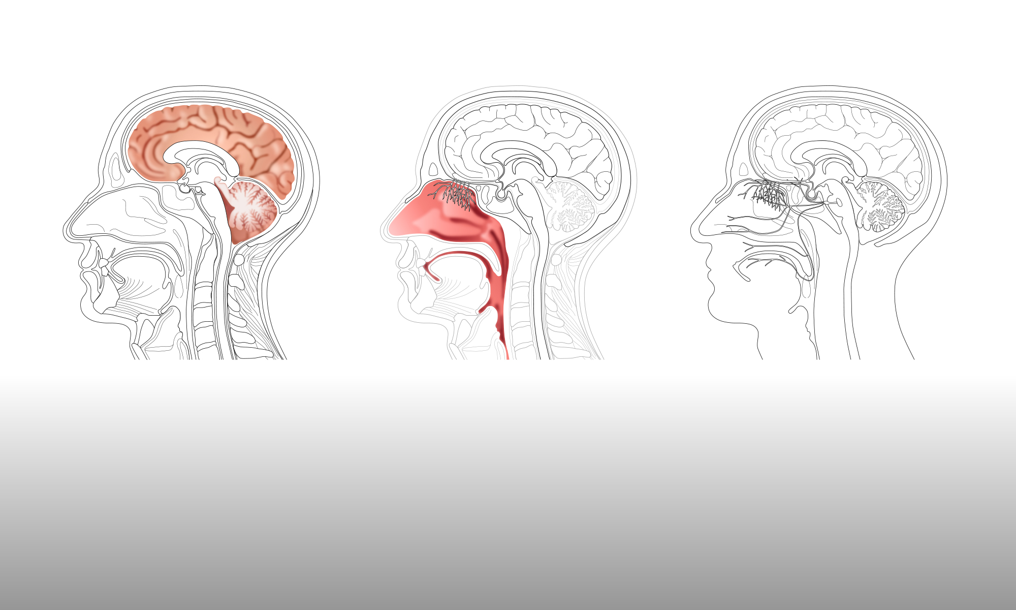
Module 5 will be designed to be a standalone ‘information sheet’, explaining what happens during MRI or PET scanning. This module will not be study specific, so can be inserted into any imaging PIS. Initially, Gill will concentrate on producing images that show MRI neuroimaging, based on one of the two main PIS examples. However, the images created will be adaptable, so that they can easily be edited to show MRI scanning of other areas of the body, or PET scanning.
The next stage is for Gill to collect reference photographs that can be used when drawing the illustrations. She will also be reviewing the images that are already in the CNS image library, to see if any can be adapted for use in the PIS project.

One Reply to “”