
Gill has been creating initial illustrative images for the various PIS modules, based on the possible images listed in this blog post. At this stage they are drawn in black outline only, with an all-white fill. This has been done for two reasons; so that time is not spent adding colour to an image that will not subsequently be used in the modules; and so that Giovanna and Mattia can consider what range of colours to use in the modules as a whole. Restricting the colours to a set colour palette (possibly using colours from the King’s branding colour palette) will make the images look more coherent as a whole, and as if they have been specifically designed for use in the PIS, rather than selected at random from Google image.
The images produced to date are shown in the galleries below. These do not include the ‘sampling images’ that were discussed in a previous blog post. All of the drawings are based on photographic images, but they typically use more than one photo as a reference and all then have additional editing and drawing applied. All of the drawings are made using Adobe Illustrator software and make extensive use of its Layer functionality, to make the images as adaptable and editable as possible. For a demonstration of how this works, see this blog post.
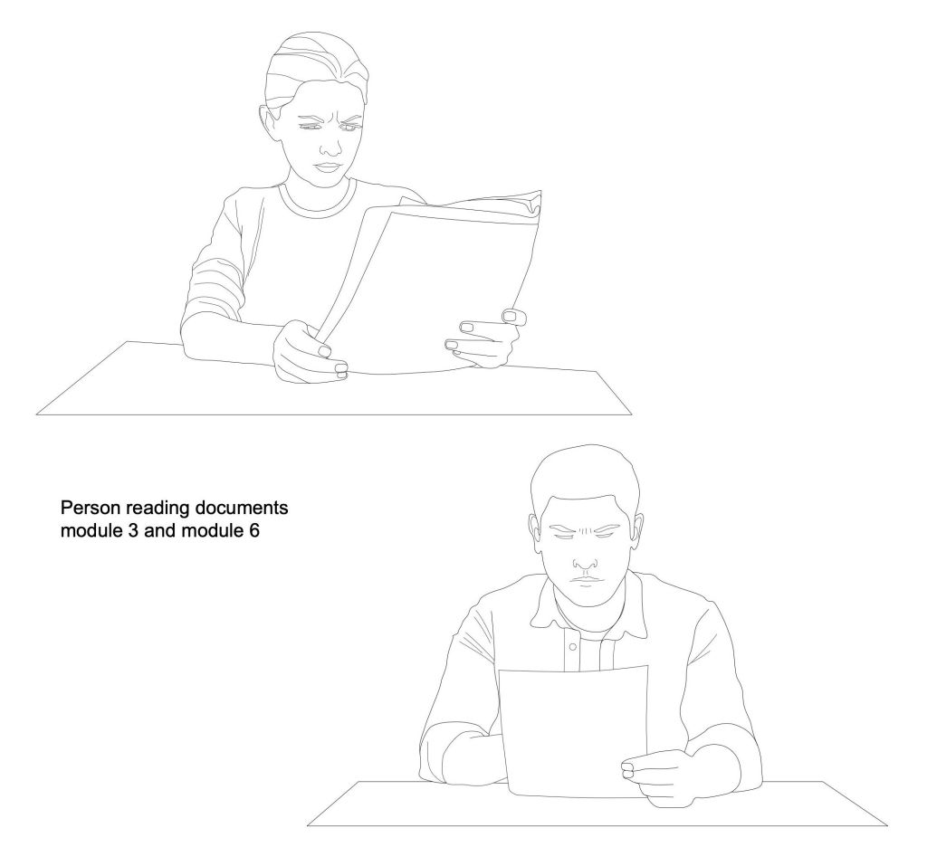
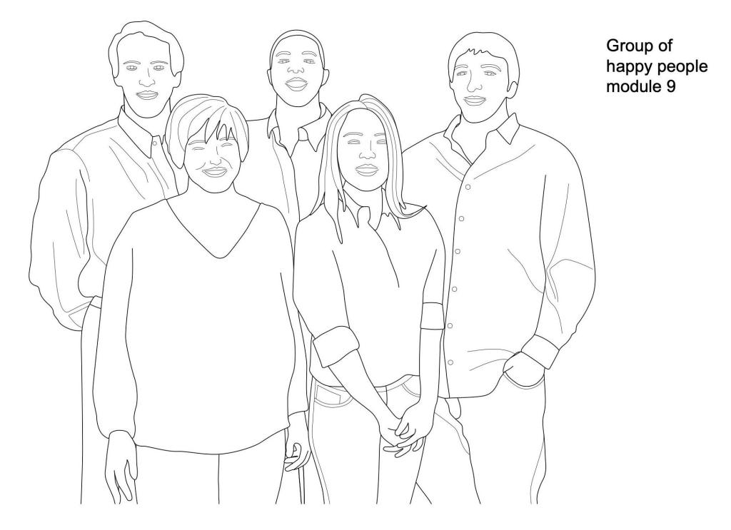
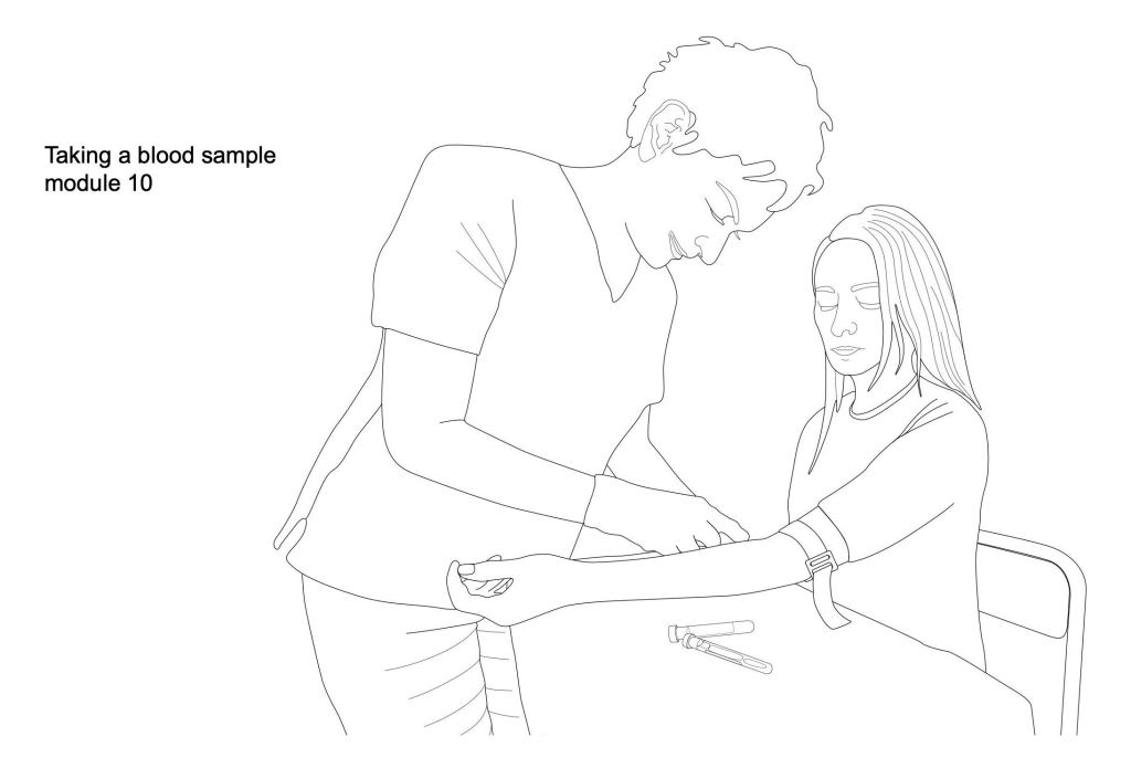
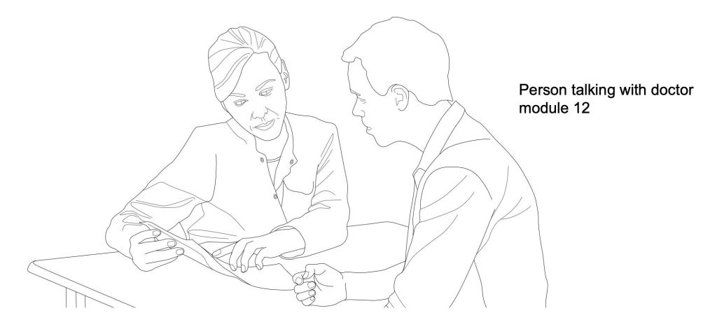
Many of the modules require images of people in various scenarios, including reading documents and talking with a clinician. The image of a clinician taking a blood sample may be used instead of an image of a syringe and needles, on the grounds it may be slightly less alarming. The images of blood sample tubes, shown in the last blog post, have been added to this image. The group of people has been constructed from individual drawings, so that people can be swapped in and out of the image, to better represent the cohort that will benefit from the imaging study.
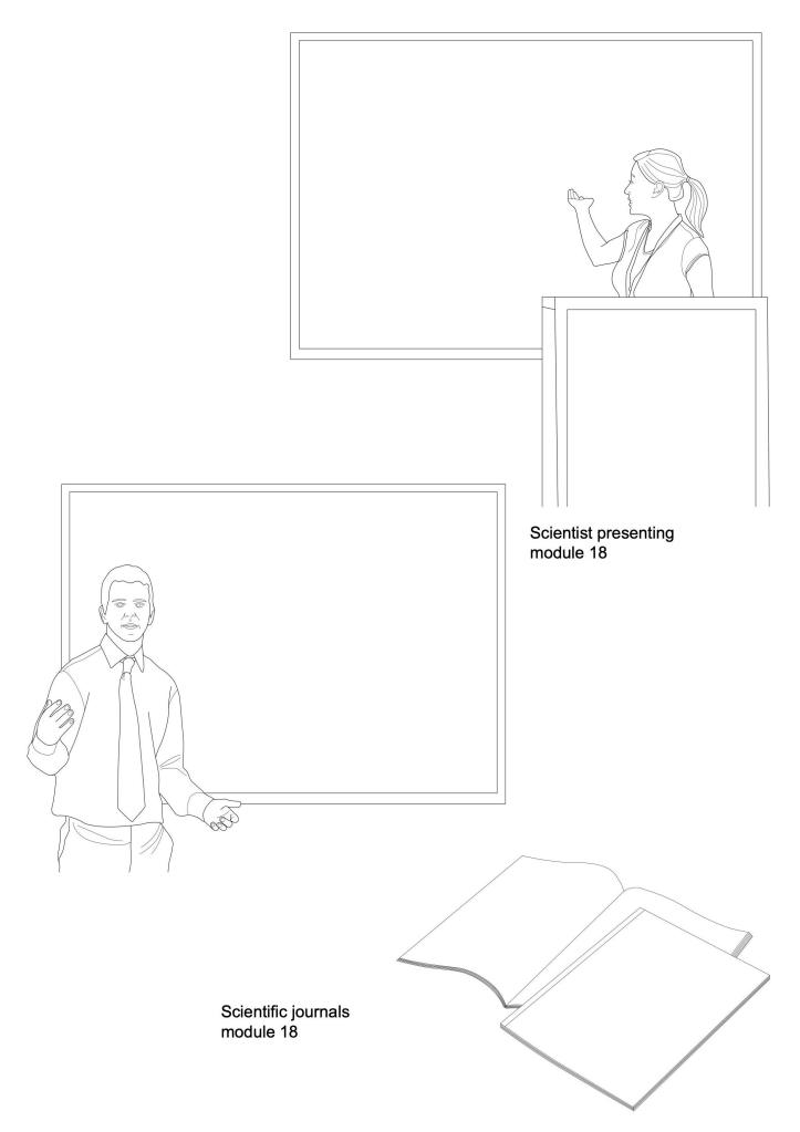
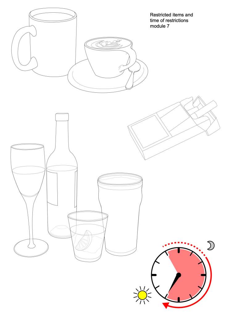
Two versions of a scientist presenting the results of a study have been drawn, together with an image of some scientific journals. The screen and page contents can be added to the images, to reflect the topic of the study. Module 7 describes those activities that may be restricted prior to taking part in an imaging study, drinking alcohol and smoking tobacco being obvious items. A drawing of E-cigarettes may also be required for this module. Showing how long the restrictions are in place is a little more complex. The image of the clock, with symbols to show night and day, will have to be adapted for each individual study and text may be needed to make the length of time absolutely clear.
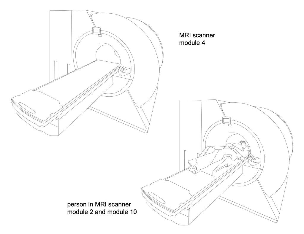
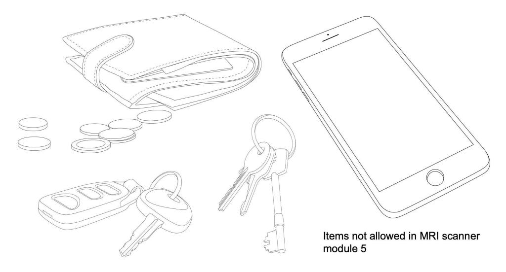
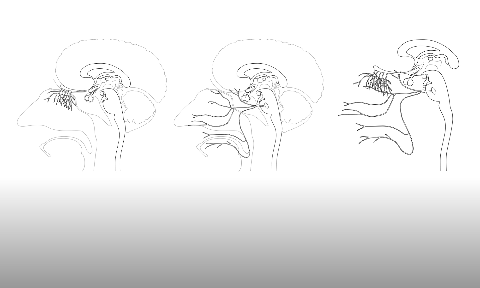
The images that still have to be drawn are those that will be needed for module 5 – the standalone guide to being scanned in an MRI scanner. However, Gill had previously created an image of an MRI scanner, with and without a patient, and you can find it in the image gallery of this website. That image could serve as a more generic representation of MRI scanning, to be used in some of the modules. The small images of items not allowed in an MRI scanner (keys, phone, etc.) could be included in module 5.
These images were sent to Giovanna and Mattia for review, with an online meeting planned for Monday 16th November to plan the next steps. In the meantime, Gill will work on the remaining images required for module 5.
