

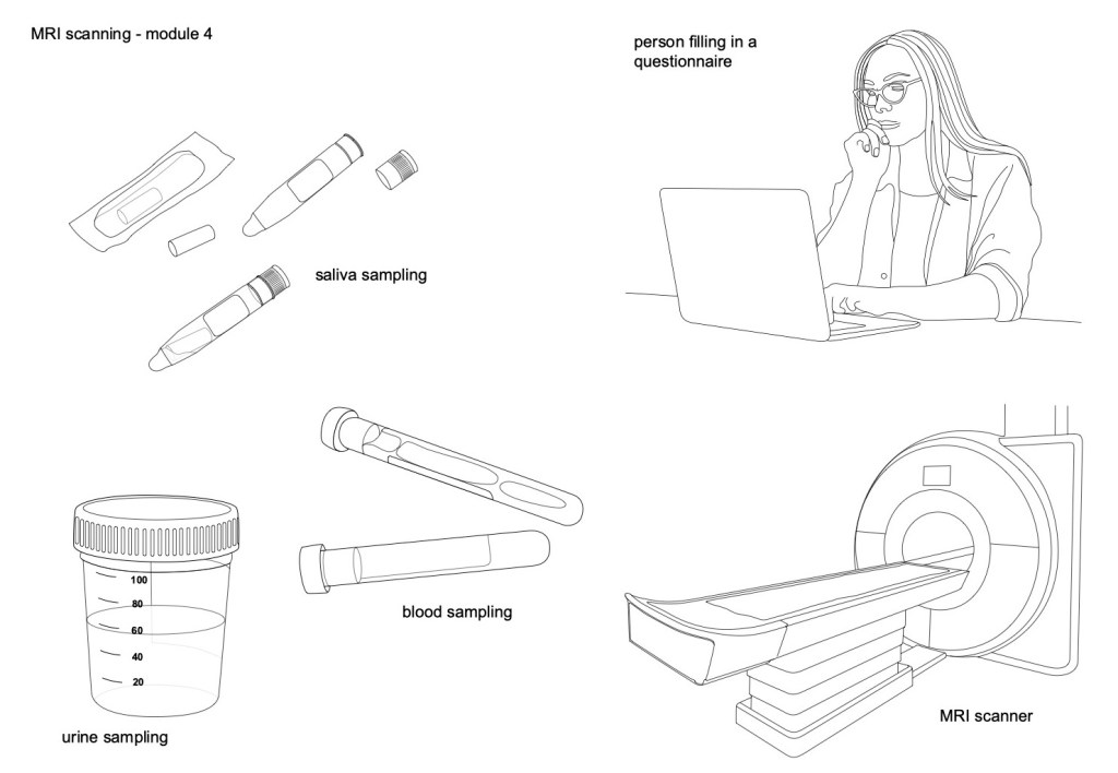
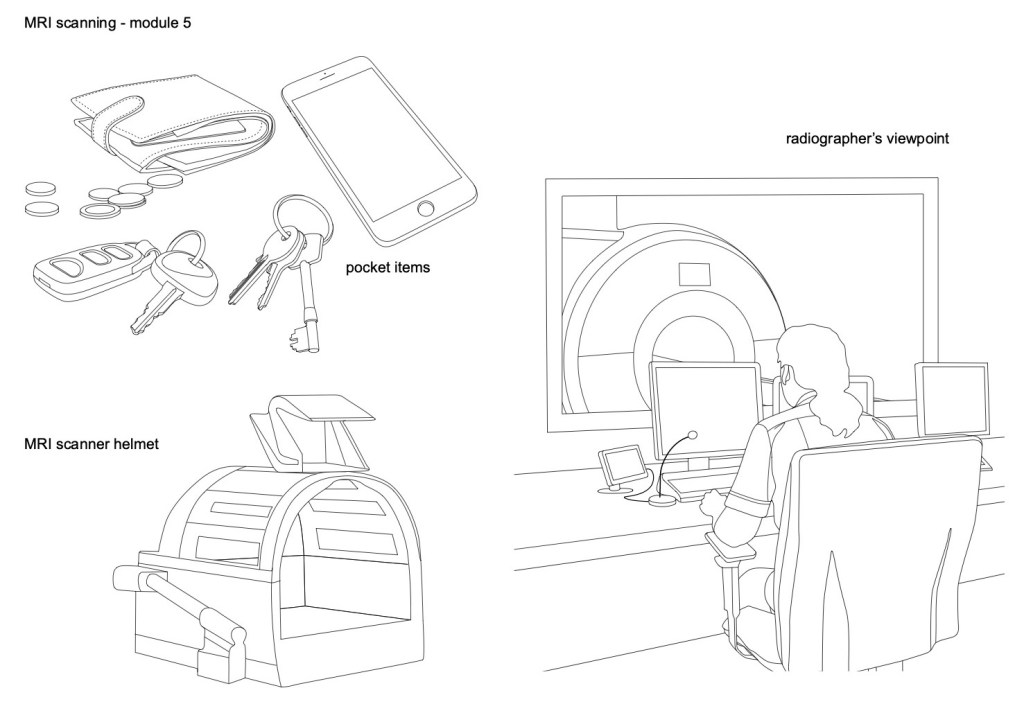
Gill has asked Giovanna and Mattia whether both Module 4 and Module 5 (rather than just Module 5) will be needed for submission to the FAST-R review, to allow a direct comparison with the MRI scanning content of the existing Patient Information Sheet. The aim of this meeting, therefore, was to review the content of the two modules and confirm the text and images that will be required for each of them. The images that have been created to date for these modules are shown below and a set of scanner images, created for possible use in module 5, are shown in the header image of this blog post.
main points from the meeting
- Modules 4 and 5 will effectively be combined into one section that will cover all of the MRI scanning content in the PIS and both will be submitted to the FAST-R. Module 4 will contain all of the information that is specific to the example PIS. Module 5 will be a more general ‘what happens during a neuroimaging MRI scan’ information document, that could be attached to any neuroimaging PIS.
- Module 4 will use the text as it is in the example PIS. This module will also include the table that summarises what happens in the two visits. In the images showing the patient and scanner in this module, the neuroimaging helmet will not be included.
- There is no explicit text relating to Module 5 in the example PIS , although some of the general MRI text can be added to the module. Giovanna and Mattia will produce the remaining text, that will describe the step by step process of a neuroimaging MRI scan. The steps will be based on the YouTube video that Giovanna had referenced at the beginning of the project, as Gill had based her choice of images on this video (rather than on any text in the PIS).
- For Module 5, Giovanna is keen to include an image that will show the inside of the scanner, to indicate to patients that it can feel like a confined space. She also wanted to draw attention to the alarm system, that allows a patient to speak to the radiographer while they are inside the scanner, as many patients have asked her about this. Gill had attempted to draw a patient’s view from inside the scanner (based on the video) but it was proving difficult to create a useful image. It was agreed that a front view of the scanner, with the patient inside, would best show the relative sizes. Gill will also add the alarm system – a ‘bulb’ that the patient can hold in their hand and squeeze to alert the radiographer – to all of the scanner images. A close-up image of a patient’s hand holding the bulb will add emphasis.
Action items from the meeting
- Gill will produce these additional images:
- A modified image of the MRI scanner, with the patient inside (with and without a helmet).
- A person signing a document, which may be used in Module 4.
- An image of an MRI scanner viewed from the front, with the patient inside to demonstrate the relatively limited space.
- A representation of the patient alarm will be added to all of the scanner images.
- A close-up view of a patient’s hand, holding the alarm. This will tie-in with the image of the radiographer at her desk with the microphone and screen showing the scanner.
- Giovanna and Mattia will produce the text specifically for use with Module 5, to describe the steps involved in having an MRI neuroimaging scan.
- Gill will create A4 templates for modules 4 and 5 , using outline images in the first instance. Once final decisions have been made, she will produce colour versions of the chosen images.
Gill will aim to have initial A4 templates for the two modules (and for Module 16) by the beginning of next week (30th November 2020).

2 Replies to “Meeting between Gill, Giovanna and Mattia, Tuesday 24th November 2020”