Skip to content

Neuroscience & graphic design

A collaboration between neuroscientists and a graphic designer

  • News
  • Projects
    • FDOPA PET Imaging Leaflet
    • Patient Information Sheet
      • Example Patient Information Sheet
    • PhD Research
  • Workshops
  • Image gallery
  • Blog
  • About the team
  • Contact

Blog

Posted on August 29, 2017October 11, 2017

Meeting between Gill, Mattia and Yannis, Friday 25th August 2017

Gill met with Mattia and Yannis to discuss the progress of the collaboration ...

Posted on August 21, 2017October 11, 2017

Human brain gallery

Creating another group of images, in order to test out a portfolio-type website ...

Posted on August 1, 2017August 2, 2017

Post 1 – Rat brain gallery

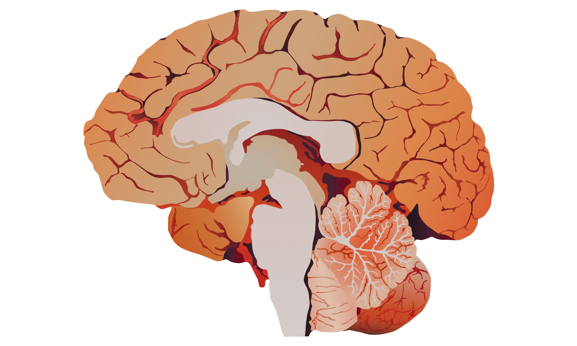
Creating a range of images of a mid-sagittal section of a rat brain, all originating from one Illustrator drawing ...

Posted on August 1, 2017August 1, 2017

Post 2 – Rat brain gallery

gallery_image9-01Aug17
gallery_image2-01Aug17
gallery_image13-01Aug17
gallery_image4-01Aug17
gallery_image5-01Aug17
gallery_image1-01Aug17
gallery_image12-01Aug17
gallery_image14-01Aug17
gallery_image6-01Aug17
gallery_image11-01Aug17
gallery_image15-01Aug17
gallery_image10-01Aug17
gallery_image8-01Aug17
gallery_image3-01Aug17
gallery_image7-01Aug17
Posted on July 26, 2017

A note on Edward Tufte

Edward Tufte is a statistician, who has written extensively on information design ...

Posted on July 24, 2017July 30, 2017

Meeting between Gill, Mattia and Yannis, Friday 21st July 2017

Gill met with Mattia and Yannis to discuss the next steps for the collaboration ...

Posted on July 17, 2017

From anatomical to diagrammatic (continued)

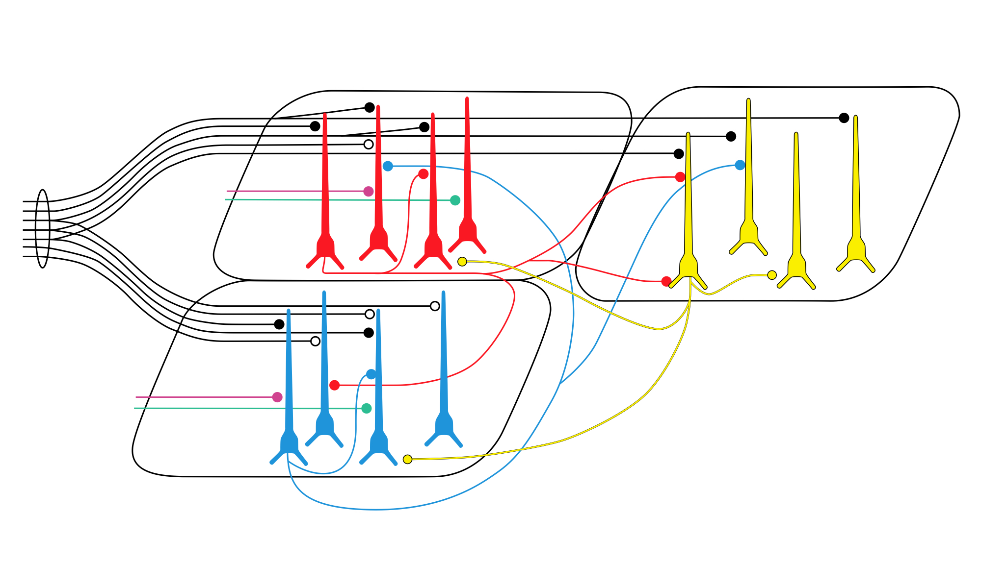
Demonstrating the range of representations for more of the visual elements ...

Posted on July 10, 2017

From anatomical to diagrammatic

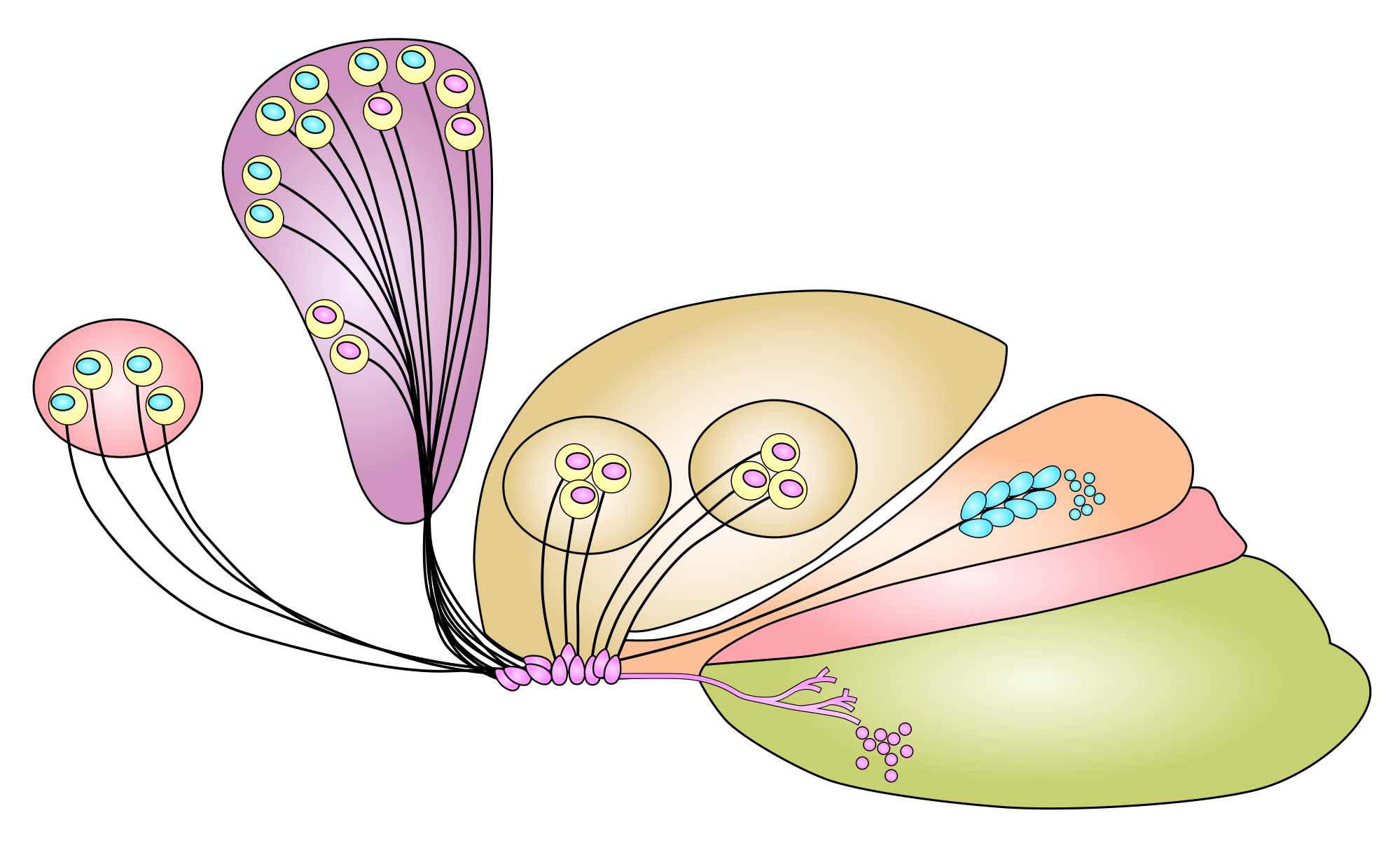
Creating a continuum of conceptual figures from anatomical to diagrammatic ...

Posted on July 6, 2017

A note on Dr Robert G. Thorne

Dr Thorne, of the University of Wisconsin, explains how and why he produces his own conceptual figures ...

Posted on July 3, 2017July 5, 2017

The olfactory epithelium

Gill has been collecting images of the olfactory epithelium, to demonstrate the range of possible visual representations ...

Posts navigation

Previous page Page 1 … Page 5 Page 6 Page 7 Next page

Go to this webpage to find out about the @neurographical Instagram feed - more than 800 images showcasing the work of the collaboration from 2017 to 2022.

An illustrative image of a human brain, one of a series created in different colourways. ©️Gill Brown #neuroscience #graphicdesign #illustration #humanbrain Re-drawn detail of a figure showing a neuron. Slightly edited to not include a big ‘drop shadow’ as I just couldn’t bring myself to draw it. I do love the overall shape, though! 😊 #neuroscience #graphicdesign #illustration #neuron An illustration showing an air traffic controller, created for use in an information sheet explaining executive function. ©️Gill Brown #neuroscience #graphicdesign #illustration #executivefunction #airtrafficcontroller
Create a website or blog at WordPress.com
  • Follow Following
    • Neuroscience & graphic design
    • Join 25 other followers
    • Already have a WordPress.com account? Log in now.
    • Neuroscience & graphic design
    • Customize
    • Follow Following
    • Sign up
    • Log in
    • Report this content
    • View site in Reader
    • Manage subscriptions
    • Collapse this bar
 

Loading Comments...