

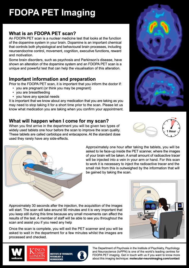
Before the meeting, Gill had produced a revised version of the FDOPA PET Imaging, changing the illustrations of the PET scans so that they had a rainbow colour scheme instead of the blue-green colour scheme used in the second version of the leaflet.
Gill had also altered the layout to include a black border around the whole leaflet, to make it more obvious that the leaflet consisted of only one page, but also to try and better integrate the black area behind the PET scans with the remainder of the leaflet. This version of the leaflet (shown left) was sent to Giovanna for review before the meeting.
At the meeting, the following points were discussed, with action items in italics:
- Giovanna liked the border around the leaflet but not the black area beneath the PET scans. She asked whether this could be replaced by just adding a black border to the scans illustrations and leaving the remainder of the background white. Gill agreed that the black area wasn’t working and she would revise the PET scan illustrations accordingly.
- Gill noted that removing the black area behind the scans meant that the black title bar could also be removed, which should provide more space for the text. Also the teat headings could be coloured (the same dark blue as the IoPPN logo), and the border could also be changed to this colour. Gill will revise the layout accordingly to see how this looks.
- Giovanna also mentioned a minor change to the text next to the logos, but otherwise the text and remaining images were fine as they were.

Gill made the changes as discussed during the meeting and produced a revised version of the leaflet (shown left) that she sent on to Giovanna and Mattia.
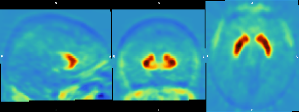
Giovanna liked the revised leaflet but had one more request – could the PET scan of the sagittal brain view (the lower PET scan illustration) show a red signal in the area of the striatum. Giovanna sent an example image (below) to show how this might look.
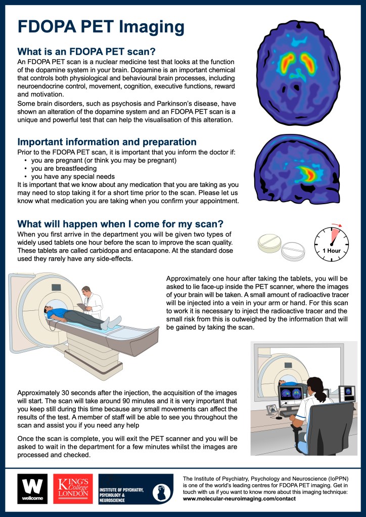
Gill revised the existing PET scan illustration to include the red signal, as shown in the header image to this blog post. This illustration was then added to the leaflet and also to the screen in the illustration of the radiographer’s office, before the updated leaflet (shown left) was sent on to Giovanna.
Giovanna was happy with the revised PET scan image, and with the leaflet as a whole. The intention now is to pass this example leaflet on to a wider group of clinicians, to gather feedback before any other versions are produced. The example leaflet describes a dynamic PET scan and, once this has been approved, a version describing a static PET scan will also be produced.
