

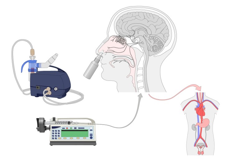
Following the meeting with Yannis, described in this blog post, Gill drew initial examples of the five visual elements that are required to demonstrate the intranasal and intravenous administration of oxytocin. These elements are shown in the header image to this blog post and are:
- Intranasal nebuliser, with nasal inhaler
- Intravenous infuser, with syringe and cannula
- Hand-held nasal inhaler
- Anatomical human head, showing nasal cavity, olfactory and trigeminal nerves and the brain
- Anatomical human torso, showing heart, stomach and reproductive organs
Drawings of the various pieces of laboratory equipment are relatively straightforward. However, Gill ensured that they were constructed in Adobe Illustrator in such a way that the equipment can be dismantled, to a limited extent, as shown below. Three different versions of the hand-held nasal inhaler were drawn, and each has a transparent cap that can be removed from the image without affecting the main bottle. Similarly, the nasal inhaler can be separated from the intranasal nebuliser, and the syringe and cannula can be removed from the intravenous infuser. This should make these files more readily adaptable for future use in other figures.

In preparation for the upcoming meeting with Yannis and Daniel, Gill constructed an initial figure to use as a starting point (shown below). This initial figure contains no text and only preliminary annotation, as the plan is for Yannis and Daniel to construct the figure for themselves during the meeting and to add the text and annotation that they require.
 The image of the anatomical head was adapted from Illustrator files that had been initially created last autumn. How the original anatomical head image was constructed is described in this blog post and the olfactory and trigeminal nerves were added later, when the images were used in the pilot workshops. The nasal inhaler, without its cap, was placed into the anatomical head Illustrator file and some additional edits were then made to make it appear as if the nozzle of the inhaler has been inserted into the nostril.
The colour image of the anatomical torso was adapted from a torso that had been drawn to contain most of the major organs, including both male and female reproductive organs, as shown below.
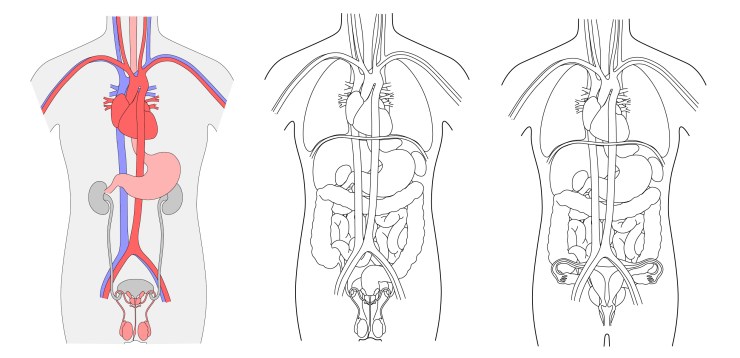
The creation of the main anatomical torso image is described in more detail in this blog post.
In the case of this figure, the torso required, as a minimum, the heart, stomach and reproductive organs. As the paper is describing experimental work carried out exclusively on male volunteers, male reproductive organs were included. However, Gill also drew female reproductive organs, so that a ‘female’ torso would be available for future figures, as shown above right.
Once the organs have been selected, there will almost certainly need to be some additional editing to ensure they appear in the right location and in the correct relationship with each other. The colour image shown above left has been edited to show the artery passing behind the stomach and to intertwine the urinary and reproductive systems. However, as Gill is definitely not an expert on human anatomy, all of this will have to be verified by Yannis and Daniel.
All of the Adobe Illustrator files for the images shown here will be provided to Yannis and Daniel prior to a meeting scheduled for 16th July 2018, where they will go through the construction of the figure. Gill will also provide them with an updated guide to how to edit these files in Illustrator, particularly the use of layers. The outcome of that meeting will be described in a future blog post.
The files for the intravenous infuser and intranasal nebuliser have already been added to the online image library and can be found in this image gallery. Versions of the anatomical head can be found in this image gallery. The hand-held nasal inhalers and the anatomical torsos will be added to the library in due course.

2 Replies to “Visual elements for an oxytocin administration figure”