

Gill met with Barbara and Mattia to discuss the conceptual figure they will need for their upcoming journal paper. The paper will review the topic of dopamine synthesis and demonstrate how this can be measured using the 18F-DOPA radioactive tracer in PET imaging. They would like one conceptual figure that shows all of the steps in the process of dopamine synthesis and how it can be measured.
There are several key papers in this field, two of which have conceptual figures that, between them, contain all of the details that Barbara and Mattia would like to include in their own figure. Both papers were published twenty years ago, so this subject has been represented visually many times, in a variety of ways.
The two figures are shown below:
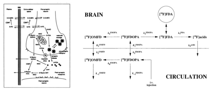
The figure on the left contains all of the visual elements that the new figure would need, although the design, and small size (only one column width in an A4 double column spread), of the figure make it quite difficult to read. The figure on the right is a purely diagrammatic representation and contains some text details that should be included in a new figure. But Barbara and Mattia both want to include the more anatomical visual elements to give the figure some context.
The header image for this blog post shows the three visual elements that will need to be included in the new figure – the blood brain barrier, neurons with a synapse (showing chemical transmission across that synapse) and dopamine receptors. The representations in the header image are taken from a figure that Barbara drew in PowerPoint, to use in a presentation on the subject, and she in turn based the drawing on a similar figure published in the journal Nature Neuroscience in 2006. Barbara and Mattia would like to create the ‘ultimate’ dopamine synthesis figure, that contains all of the information that has been included, in one way or another, in the figures created over the preceding 20 years. Gill’s aim is to produce an ultimate figure that can then be easily edited and adapted for a range of different circumstances, depending on how and where the figure is to be used.
The first stage in the process is for Gill to collect examples of the three visual elements. She is familiar with visual representations of chemical transmission across a synapse, as that has been a key image in her PhD research. But the representations of the blood brain barrier and of dopamine receptors (as opposed to other types of receptors) will be new visual elements for this collaboration. As they are collected, examples of all three elements will appear on the neurographical Instagram feed.
Once Gill has a feel for how these elements should appear, she will draw examples that can be used in the new figure and have a first attempt at creating the figure as a whole. The figure, and elements, will be drawn in Adobe Illustrator and will utilise the Layer functionality to make building the figure as simple, and as flexible, as possible. At that stage it will then be necessary to meet again with Barbara and Mattia, so that they can work together with Gill to construct the figure as they want it, and to understand how to edit it. The whole process will be documented in future blog posts.
The journal papers discussed during this meeting were:
Cumming, P. and Gjedde, A. (1998) Compartmental Analysis of Dopa Decarboxylation in Living Brain from Dynamic Positron Emission Tomograms. Synapse 29, pp37-61.
Holden, J.E. et al (1997) Graphical Analysis of 6-Fluoro-L-Dopa Trapping: Effect of Inhibition of Catechol-O-Methyltransferase. The Journal of Nuclear Medicine, Vol. 38, No. 10, pp 1568-74.
Huang, S-c. et al (1991) Kinetics and Modeling of L-6-[18F]Fluoro-DOPA in Human Positron Emission Tomographic Studies. Journal of Cerebral Blood Flow and Metabolism, Vol. 11, No. 6, pp. 898-913.

3 Replies to “Meeting between Gill, Barbara and Mattia, Wednesday 6th June 2018”