
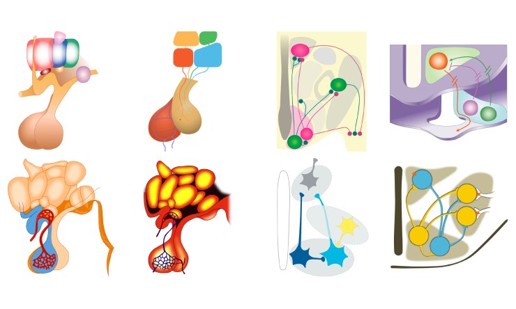
Since July 2017, Gill has been collecting examples of the visual elements that would be required for the conceptual figures in Yannis and Nisha’s journal articles. Gill has re-drawn the elements in Adobe Illustrator, to remove them from the context provided by their original conceptual figures, and has excluded any text and most of the annotation. The resulting images have been posted on the neurographical Instagram feed, with over 150 examples now added. Some of these images are shown in the header image.
For a recent presentation about her research, given to post-graduate graphic design students at London College of Communication, Gill collected together the re-drawn elements into categories, to see what conclusions could be drawn simply from their physical appearance. It is worth recording these interim conclusions in this blog post.
Seen all together, the general appearance of all of the elements is very varied, particularly in the colours that are used. However, once they are separated into their groups, similarities become more obvious. Not all of the examples that are collected on Instagram are shown here, but these are representative samples.
It is probably easiest to start with the most familiar examples, the human brains:
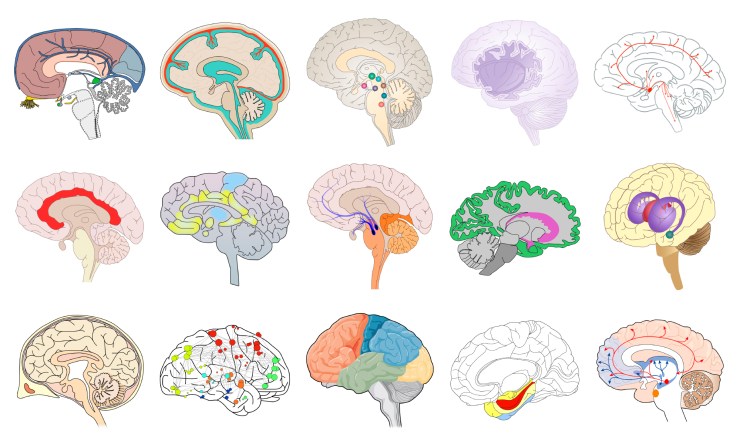
A sagittal section through the brain is the most common representation and the vast majority are shown with the front of the brain on the left (the same is true for figures of the human head). Colours can be extremely variable, but the shapes used are very similar and the same regions are often highlighted – the cerebellum, corpus callosum, cortex and brainstem.
Rat brains continue in a similar vein, with the sagittal section being most common, and the front of the brain almost always shown on the left.
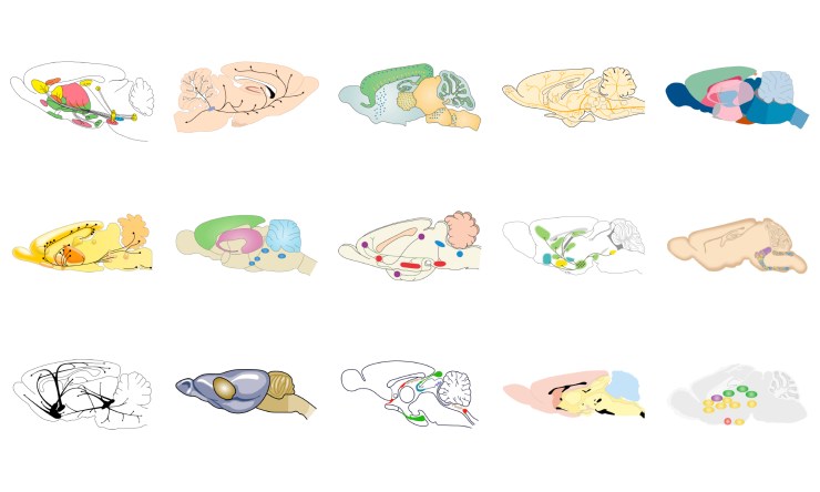
The shape of the brain is a little more variable, possibly because it is not as familiar, but the large olfactory bulb at the front of the brain, and very distinct cerebellum at the rear, are almost always highlighted.
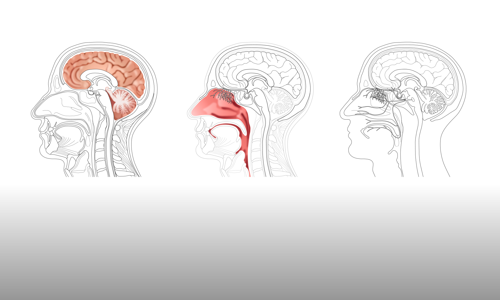
Human nasal cavities and olfactory nerves are usually shown in the context, with at least part of the head, skull and/or brain included in the figure.
 Again, as with the brains, the images almost all face to the left. One style of representation is most prevalent, as shown in the centre and right of the top row, with the olfactory bulb of the brain at the top of the nasal cavity.
The representation of the hypothalamic nuclei fall into three main groupings, with the first two shown below:
The more anatomical representations, which always include the pituitary gland, are shown on the left. The pituitary gland is often shown with its blood supply, and the nuclei are represented by rounded, or geometric shapes. On the right, the representations are much more diagrammatic, with more emphasis on the functions of the hypothalamic nuclei and how they connect with each other.
The third style of representations, shown below, could be termed the ‘dancing pituitaries’, with the nuclei shown either as geometric shapes, or as large ‘neurons’ that connect with the pituitary gland.
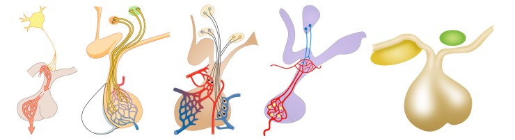
Putting the colours to one side, the shapes used in all three representations are very similar within each group and very recognisable.
Representations of brainstem nuclei also fall into three broad groups – side view, front view and cross sections – as shown below.
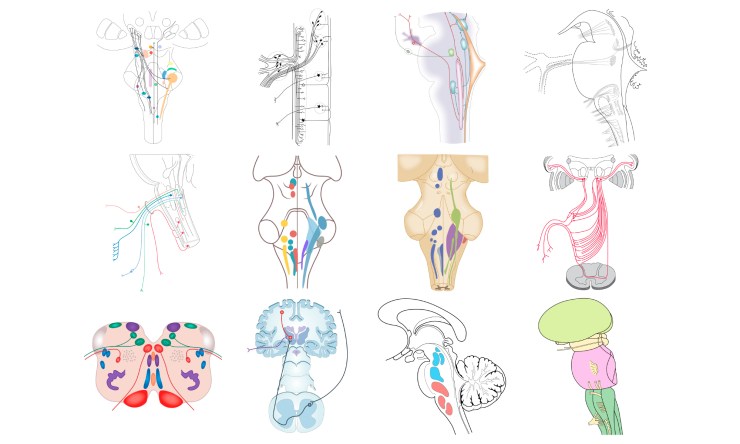
The nuclei themselves are represented either by geometric shapes or, if connections between the nuclei are important, are shown as connecting lines, in a similar style to a circuit diagram.
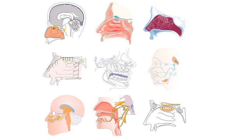
The olfactory epitheliums show the most variation in their appearance, as shown below.
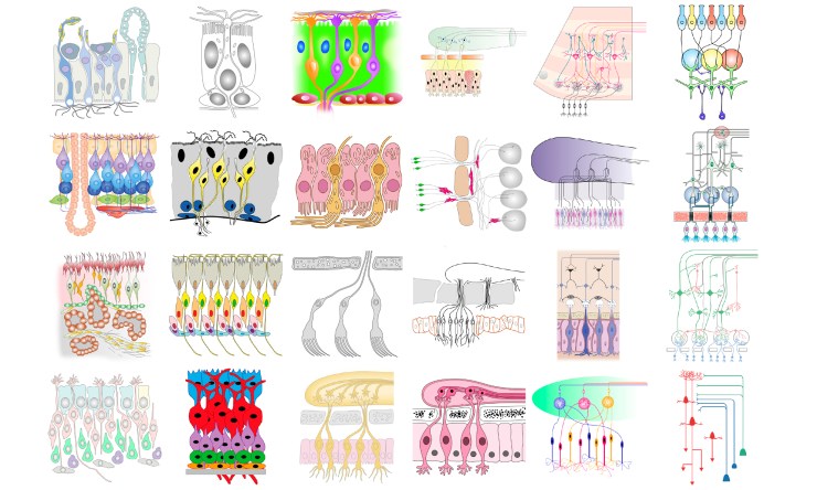
The most anatomical representations are shown on the top left and these become more diagrammatic towards the right, with the most diagrammatic representation at the bottom right. The images can also be divided into those that include the olfactory bulb and olfactory nerves, and those that show only the epithelium. The choice of representation presumably depends on the context of the figure and what is being emphasised in the journal article – either the anatomy of the epithelium or the connections formed by the olfactory nerves.
Lastly, the collections of neurons are shown below:
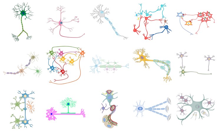
Although, once again, the colours are very variable, the shapes used to represent a typical neuron are well-established, with the axon and dendrites present in every case. The amount of detail, in terms of depicting the myelin sheath, or nerve cell nucleus, etc., does vary, presumably depending on context.
Collecting a large number of visual elements, and drawing them for herself, has given Gill a very good understanding of which visual aspects must be included in an element in order for it to be instantly recognised by a viewer. Equally, she now knows how much leeway exists for variation and individuality when creating these elements for use in conceptual figures.
